
X-rays are an essential band of the electromagnetic spectrum. Since Wilhelm Conrad Röntgen discovered them in 1895, X-rays have transformed healthcare, material science, and global security. Understanding their fundamental physical, chemical, and biological properties explains how they penetrate solid matter and how to safely control them.
[IMAGE PLACEHOLDER: Historical photo of Wilhelm Röntgen alongside a modern high-resolution digital X-ray image.]
1. Fundamental Electromagnetic Characteristics
- Electromagnetic Nature: X-rays consist of massless energy packets called photons traveling in a vacuum at the speed of light (c≈3×10⁸ m/s).
- Zero Electric Charge: X-ray photons carry no electric charge. Consequently, external electric or magnetic fields cannot deflect or bend an X-ray beam.
- Wave-Particle Duality:
- Wave Behavior: They display wave properties such as diffraction, interference, and polarization at atomic dimensions.
- Particle Behavior: At high energies, they exhibit particle properties through momentum transfer, as seen in the Compton effect and Photoelectric effect.
- Spectrum Position: X-rays sit between ultraviolet radiation and gamma rays. Their wavelengths span 0.01 nm to 10 nm, corresponding to frequencies between 3×10¹⁶ Hz and 3×10¹⁹ Hz.
2. Mechanics of X-Ray Generation
X-rays are generated inside a vacuum tube where high-speed electrons collide with a dense metallic target (typically tungsten or molybdenum).
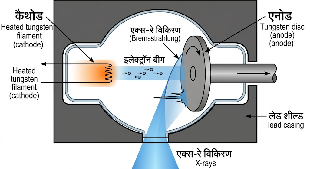
- Continuous Radiation (Bremsstrahlung): When fast-moving electrons pass near heavy target nuclei, the intense electrostatic field slows them down. This loss of kinetic energy is released as continuous X-ray photons.
- Characteristic Radiation: When an incoming electron knocks out an inner-shell electron of the target atom, an electron from a higher shell drops down to fill the gap. This transition emits an X-ray photon with a precise energy characteristic of that specific element.
3. Core Physical Properties & Matter Interactions
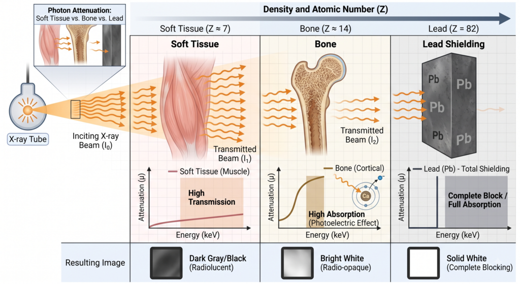
- Penetration & Exponential Attenuation: X-rays penetrate opaque materials. As an X-ray beam travels through matter, its intensity decreases exponentially according to Beer-Lambert’s Law:I=I0e−μx(Where I is final intensity, I0 is initial intensity, μ is the linear attenuation coefficient, and x is material thickness).
- Atomic Number Dependency: Attenuation depends heavily on a material’s atomic number (Z) and density. Materials with high Z numbers (e.g., Lead, Z=82) absorb X-rays far more effectively than low Z elements (e.g., Hydrogen or Carbon in soft human tissue).
- Ionization Capability: X-rays carry enough energy to knock tightly bound electrons out of atoms, creating positive ions and free electrons.
- Fluorescence and Phosphorescence: Striking compounds like zinc sulfide (ZnS) or barium platinocyanide converts invisible X-ray energy directly into visible light.
4. Chemical and Biological Properties
- Photographic Effect: X-rays expose photographic emulsions and digital flat-panel detectors similarly to light, turning exposed silver halide crystals dark.
- Chemical Action: X-ray radiation can break chemical bonds, trigger free radical formation, and catalyze polymerization or color changes in specific solutions.
- Biological Effect:
- Direct Damage: Directly breaks double-stranded DNA chains inside cell nuclei.
- Indirect Damage: Radiolyzes water molecules within cells (H2O→H++OH−), creating free radicals that damage cellular structures.
- Therapeutic Utility: Controlled high doses destroy malignant tumor cells (Radiation Therapy).
5. Detailed Classification: Soft vs. Hard X-Rays
| Property | Soft X-Rays | Hard X-Rays |
|---|---|---|
| Wavelength | 0.1 nm to 10 nm | 0.01 nm to 0.1 nm |
| Photon Energy | 100 eV to 10 keV | 10 keV to 100+ keV |
| Penetration Depth | Low (absorbed by thin air or paper) | High (penetrates dense metals and human tissue) |
| Primary Uses | Surface analysis, soft-tissue microscopy | Radiography, CT scans, industrial inspection |
6. Comprehensive Real-World Applications
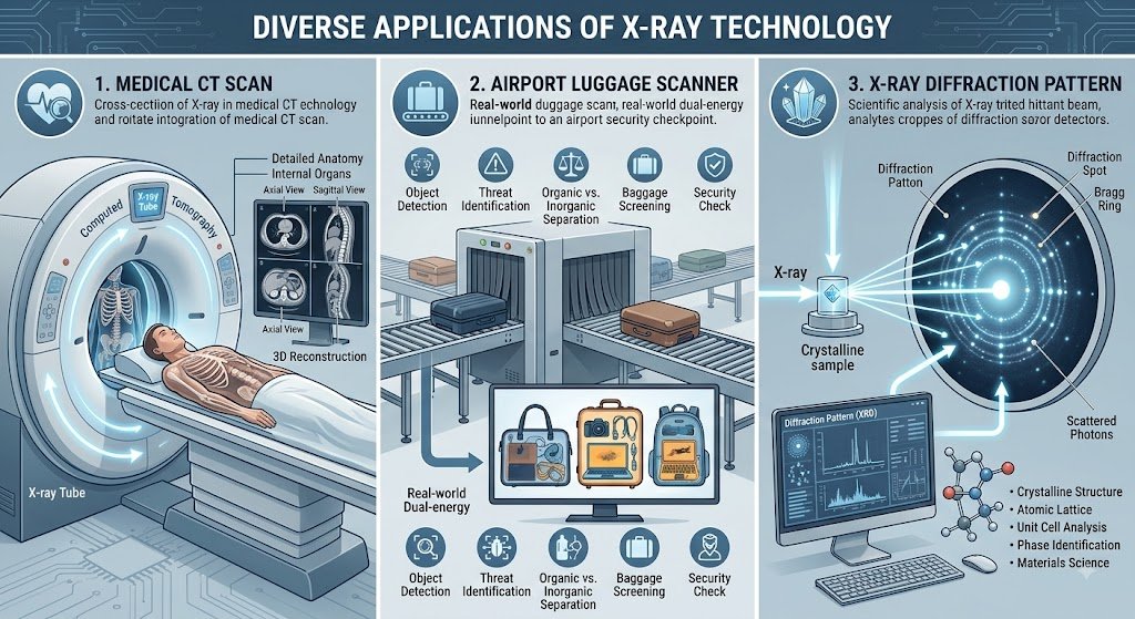
- Medicine & Diagnostics:
- Radiography: Detects bone fractures and dental issues.
- Computed Tomography (CT): Combines multiple cross-sectional X-ray images into detailed 3D models.
- Fluoroscopy: Real-time moving X-ray images for guiding surgical procedures.
- Industrial & Security:
- Non-Destructive Testing (NDT): Inspects internal structural cracks in airplane wings, pipelines, and concrete without damaging the object.
- Cargo & Security Scanners: Identifies concealed weapons, explosives, and contraband at transit hubs.
- Scientific Research:
- X-Ray Crystallography (XRD): Analyzes molecular crystal lattices to determine the 3D structure of complex proteins, DNA, and advanced materials.
- X-Ray Astronomy: Detects high-energy emissions from black holes, neutron stars, and supernova remnants using orbital telescopes.
7. Safety Protocols & Protection Principles
Because X-rays are ionizing radiation, safety protocols follow the ALARA principle (As Low As Reasonably Achievable):
- Time: Minimize the duration of exposure.
- Distance: Increase distance from the radiation source (intensity drops following the inverse-square law: I∝r21).
- Shielding: Place dense materials like lead aprons, leaded glass, or concrete barriers between the source and humans.
0 responses on "The Ultimate Guide to X-Ray Physics: Comprehensive Properties, Generation, and Modern Applications"